")
")

Cardiac MRI
It is based on the use of a strong magnetic field and radio waves to synthesise images of the heart and chest, while the images obtained are processed using specialised software installed on a computer. Cardiac MRI is one of the most important imaging techniques for the diagnosis of cardiovascular disease. Most often, during the examination, an intravenous contrast agent is administered to obtain additional information from the images.
What are the advantages of Cardiac MRI?
It offers high-resolution images and allows for three-dimensional imaging of the heart chambers and chest vessels. Τhe contrast agent used during the exam has a lower risk of causing an allergic reaction than contrast agents used in other imaging tests and MRI of the heart is a non-invasive, bloodless imaging test that does not expose the patient to ionizing radiation.
How is the test performed?
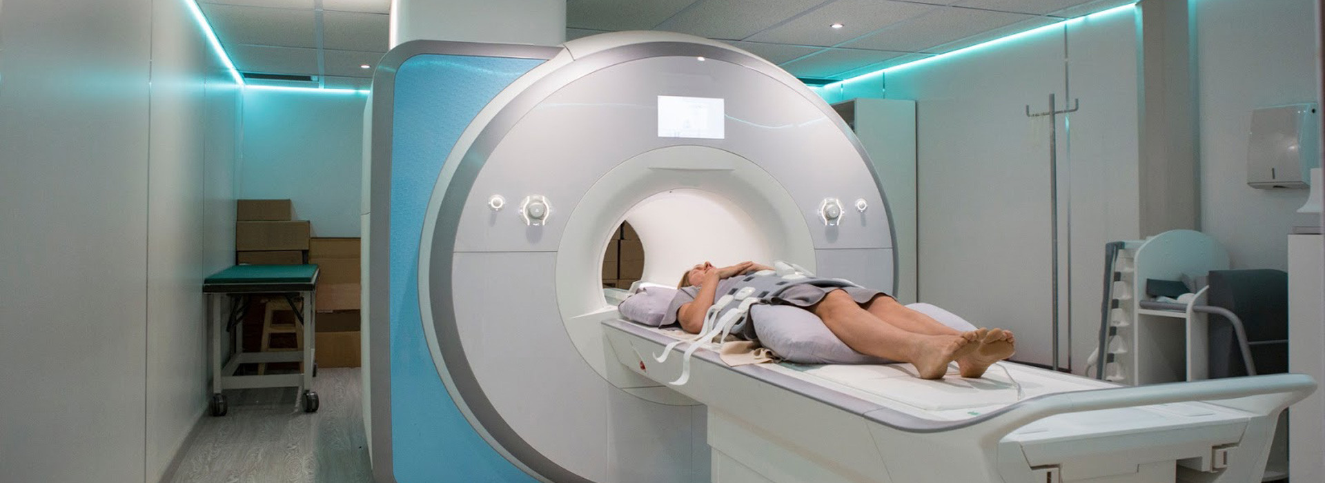
The patient lies in a supine position (on his/her back) on the special bed table of the scanner. We place 3 stickers on the chest for continuous monitoring of the heart rate. The patient wears special headphones to follow the operator's instructions. If contrast agent is required, a venipuncture catheter is placed by the nurse to administer the drug intravenously about halfway through the examination. The examination takes a total of approximately 30-45 minutes depending on the indication and findings. After the examination there is no restriction, the examinee can eat or drink freely and there is no question of avoiding contact with children as no radiation-emitting formulation has been administered.