")
")

Coronary Arteriography
The diagnosis and treatment of coronary artery disease require accurate imaging of the coronary arteries. If there is evidence that the coronary arteries have narrowings or blockages, the most accurate visualization of the arteries and branches is done with Coronary Arteriography.
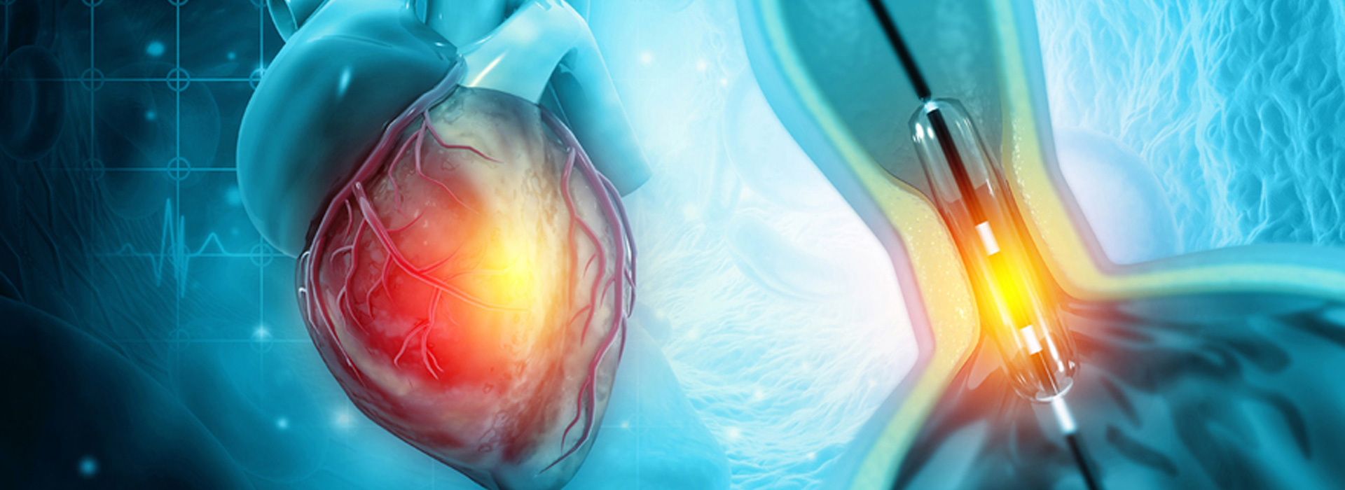
It is a minimally invasive radiopaque imaging technique of the coronary arteries after injection of a radiographic contrast medium through special catheters. Coronary Angiography is performed underlocal anesthesia through puncture of a superficial artery under the skin, then a thin flexible tube (cardiac catheter) is inserted into the artery and advanced to the heart. Through the cardiac catheter, a contrast agent is injected, while at the same time the course of the heart's arteries is recorded from different angles, thus revealing any abnormalities. During the examination, the patient's heart rate and vital signs are constantly monitored.
The arteries used for puncture and insertion of cardiac catheters are the femoral artery in the groin (i.e. the upper thigh), the brachial artery which is in the flexor surface of the elbow, and the radial artery in the forearm (wrist). In the past, the femoral artery was mainly used, while now, the radial artery (radial access) is mostly used. The major advantages of radial access are the minimization of bleeding complications as well as the immediate mobilization of the patient.
To visualize the coronary vessels, a catheter must be inserted, one end of which will reach the coronary arteries, while the other end is handled by the interventional cardiologist. To insert the catheter, a peripheral artery (an artery distant from the heart) is punctured. The catheter is then inserted and advanced to the ostia of the coronary arteries. In this position, the contrast medium is administered, which, due to its properties, provides imaging of the lumen of the vessels and not of the heart or other anatomical structures.
The images are taken in various views, moving the examination table and/or the X-ray machine, to obtain the necessary information, by imaging the arteries along their entire length. By using newer techniques (IVUS, OCT), it is possible to insert special catheters that provide data on the thickness, composition of the atherosclerotic plaque, the presence of rupture, etc. Also, the catheter can be advanced into the left ventricle and information about the heart's function, as well as the function of the valves, can be obtained.
After the examination, which usually lasts 15-30 minutes, we draw conclusions about the treatment that the patient is recommended to follow (pharmaceutical, angioplasty, surgery). The patient's stay in the hospital ranges from a few hours to a day depending on the judgment of the doctor.